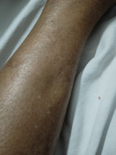
A case of lower limb weakness and Edema
Hello everyone, this is a case discussion of a 18year old male who came to the OPD with Chief complaints of bilateral lower limb weakness and edema.
- I've been given this case to solve in an attempt to understand the topic of "patient clinical data analysis" to develop my competency in reading and comprehending clinical data including history, clinical findings, investigations and come up with a diagnosis and treatment plan.
You can find the entire real patient clinical problem in this link here made by our interns
https://hitesh116.blogspot.com/2020/05/12may-2020-elog-medicine-intern.html?m=1
https://srianugna.blogspot.com/2020/05/hello-everyone.html
FOLLOWING IS MY VIEW ON THE CASE-
Chief complaints according to my order of priority-
1-Bilateral lower limb weakness-20 days
2-bilateral lower limb edema
Now let us discuss the each complaint individually and In a detailed manner-
Nervous system problem?
Now let us discuss the each complaint individually and In a detailed manner-
Weakness -
-bilateral lower limbs
-since-20 days
-started in the proximal region 2years back
-insidious in onset
-gradually progressive
-later progressed to distal parts bilaterally
-associated with difficulty in squatting and getting up from squatting position.
-associates with wearing and holding chappals
Now, what causes weakness of bilateral lower limbs?
May be due to any pathology in the arteries or nervous system-
-from the general examination we can rule out arterial disease as there is no history of pain when walking or the skin on the legs in normal and no loss of hair on the skin which indicates the absent of peripheral artery disease and varicose veins which are one Of the main causes of Leg weakness.
GENERAL EXAMINATION-
-patient was conscious, coherent and coperative
-moderately built and nourished.
-no signs of pallor, icterus, clubbing, cyanosis, lymphadenopathy, edema
-VITALS
1.temperature-AFEBRILE
2.pulse rate-92bpm
3.respiratory rate-18 cycles/min
4.BP-130/90mmhg
5.SpO2-96%
6.GRBS-142mg/dl
SYSTEMIC EXAMINATION
I.CVS-
S1 S2 heard
no added murmurs
2.RESPIRATORY SYSTEM-
-normal vesicular breath sounds heard
-bilateral air entry present
3.PER ABDOMEN-
shape=scaphoid
umbilicus=central and normal in position
all quadrants moving equally on respiration
no tenderness
no organomegaly
bowel sounds-heard
no bruit heard
4.CNS-
patient is conscious, coherent, coperative
patient well oriented to time, place and person
higher mental functions= normal
Cranial nerves- intact
Motor system-
tone - normal
power - 4-/5 in both lower limbs
reflexes absent in both lower limbs
sensory system-normal
No meningeal signs
No cerebellar signs
-from the above complaints the patient is unable to wear his chappals properly which indicates a problem in the nerves as In a demylinating disorders like CIDP.
-CIDP also causes weakness of the muscles,gradually over 8-6 weeks of time periods and also slows down the reflex.
-from above CNS examination we can see that there is hypotonia and hyporeflexia which are the main features of Lower motor neuron lesion!
Now what are lower motor neuron lesions?
Refers the above article LMN lesions
Important signs of LMN lesions are-
-hypotonia
-hyporelfexia
-weakness (patients chief complaint)
-muscle atrophy (patients histopathology showed signs of atrophy)
-effects are limited only to a small group of muscles (as the patinet has difficulty in squatting
nd getting up form that position)
So based on the above signs and results we can say that it is related to the muscle mainly and might be a myopathy.
What is myopathy?
http://www.clevelandclinicmeded.com/medicalpubs/diseasemanagement/neurology/myopathy/
Based on the above article among the many types of myopaties it might be the inherited type muscular dystrophy from the following data-
 Creatinine levels are raised which indicates muscular atrophy and also the above muscle biopsy shows femoral atrophy , muscle weakness, edema of the legs which can be interpreted as muscle enlargement and the patients inability to squat and wear cheppals all contribute to muscular dystrophy.
- now again muscular dystrophy is many types but the most common are
-duchenne muscular dystrophy
-Becker’s muscular dystrophy
•duchenne muscular dystrophy can be ruled out as the patient cannot walk without support and usually requires a wheelchair and might not survive as well.
This makes us to conclude the differentials as-
-Becker’s muscular dystrophy
https://www.mda.org/disease/becker-muscular-dystrophy
-CIDP
The patients current treatment is-
nd getting up form that position)
So based on the above signs and results we can say that it is related to the muscle mainly and might be a myopathy.
What is myopathy?
http://www.clevelandclinicmeded.com/medicalpubs/diseasemanagement/neurology/myopathy/
Based on the above article among the many types of myopaties it might be the inherited type muscular dystrophy from the following data-
 Creatinine levels are raised which indicates muscular atrophy and also the above muscle biopsy shows femoral atrophy , muscle weakness, edema of the legs which can be interpreted as muscle enlargement and the patients inability to squat and wear cheppals all contribute to muscular dystrophy.
- now again muscular dystrophy is many types but the most common are
-duchenne muscular dystrophy
-Becker’s muscular dystrophy
•duchenne muscular dystrophy can be ruled out as the patient cannot walk without support and usually requires a wheelchair and might not survive as well.
This makes us to conclude the differentials as-
-Becker’s muscular dystrophy
https://www.mda.org/disease/becker-muscular-dystrophy
-CIDP
The patients current treatment is-
T Prednisolone 15mg po od
T Pantop 40mg bbf
T Met xl 12.5mg od
Cap Becosules od
T Chymerol forte od
T Taxim 200mg bd
T Vit c od
Treatment plan for Becker’s dystrophy and CIDP are-
-There is no cure, but treatments are available to help with symptoms and maximize muscle function. It is vital that a person with BMD stay in shape and continue to use their muscles. This can include physical therapy. Treatment can also include genetic counseling, using splints, massages, and catabolic steroids.
-Treatment for CIDP includes corticosteroids such as prednisone, which may be prescribed alone or in combination with immunosuppressant drugs. Plasmapheresis (plasma exchange) and intravenous immunoglobulin (IVIg) therapy are effective. IVIg may be used even as a first-line therapy.
References-


Comments
Post a Comment